As the last in this series on the skeletal anatomy of the hand, we are now up to the fingers. This area can be tricky as beginners generally just add the fingers to the top of the body of the hand and have not worked it out to structurally fit the skeleton.
Today, we will take a look at the finger bones to see how these bones connect the fingers to the hand, allow the fingers to move, and identify the differences between the bones of the fingers and the thumb.
Let's take a look.
The bones of the fingers and thumb
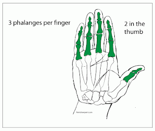
The last group of bones in the hand we are looking at are the bones of the fingers. These are called the phalanges. Each bone in this group individually is called a phalanx.
Each phalanx makes up one segment of the figure, joint to joint. Though, take note as to where the proximal phalanx starts on the hand. Looking at the palm we can see that the base of the proximal phalanx starts within the palm and not at the top.
The number of phalanges
There are three phalanges in each finger and two phalanges in the thumb. This may seem confusing when we examine the movement of the thumb as there are three bones that can be moved in the thumb. As we saw in a previous post, one of the metacarpal bones is involved in thumb movement. Whereas the metacarpals attached to the fingers do not move when we move the fingers.
Movement
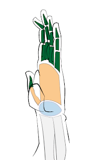
These final illustrations demonstrate the types of movements the phalanges are shaped to allow. In the first illustration, we are starting with the fingers extended.
The type of joint between each phalanx and between the phalanx and the metacarpals is a hinge joint. This gives us a hint as to the type of movement that the fingers perform, flexion and extension.
The bones are shaped at the joint to allow the phalanges to decrease in angle so that we can draw the fingers towards the palm, then extend them back to their original position.
Here is an example of a finger bending towards the palm at the base of the proximal phalanx. Remember the base of the phalanx is below the top of the flesh of the palm. When the finger bends this compresses the flesh of the palm together.
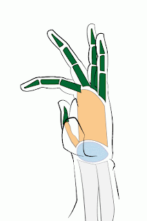
Here is an example of movement at the joint between the proximal and medial phalanges. This is the same type of joint allowing for the same kind of action. The combined movement allowed by the two joints brings the tip of the finger closer to the palm.
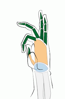
Finally, here is an example of the movement allowed by the joint between the medial and distal phalanges. The three finger bones and the three joints connecting them allow the fingers to bend completely into the palm to grip or make a fist.
To further examine this, take a look at your hand from the side while your fingers are extended. Then slowly cup your hand. As the cup forms watch where the bending occurs and note the shapes created.
To further examine this, take a look at your hand from the side while your fingers are extended. Then slowly cup your hand. As the cup forms watch where the bending occurs and note the shapes created.


